.
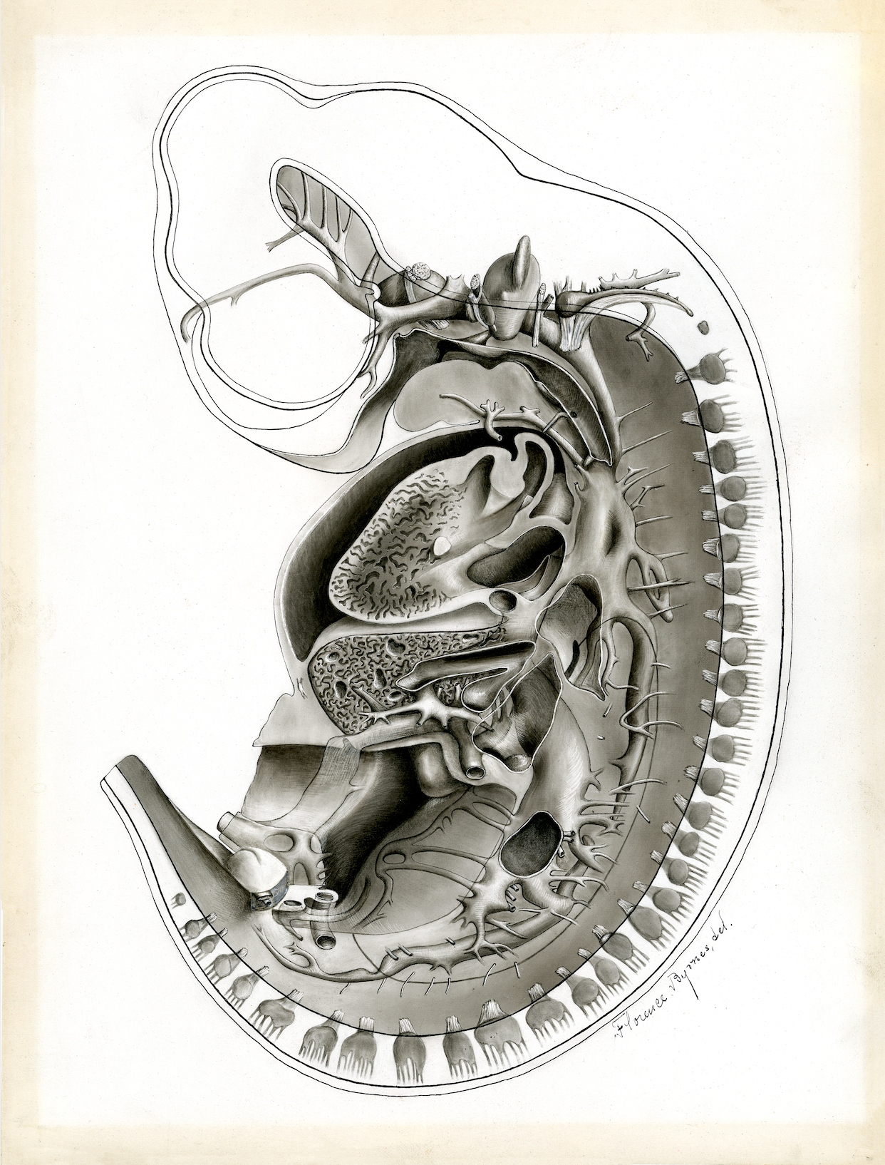
Transverse Section of Pig Embryo
This drawing is an idealized reconstruction basedon hundreds of individual sections, microscope observations, a variety of measurements, and careful observation of a wax model. It was meant to give students a better understanding of the development of an embryo than they could get from an image of any individual example. Harvard Medical School illustrator, Florence Byrnes, worked with a medical school instructor, Frederic T. Lewis, to create this image for professor Charles Sedgwick Minot’s 1903 Laboratory Textbook of Embryology.
Florence Byrnes
1903
Courtesy of Warren Anatomical Museum, Center for the History of Medicine, Francis A. Countway Library of Medicine, Harvard University