.
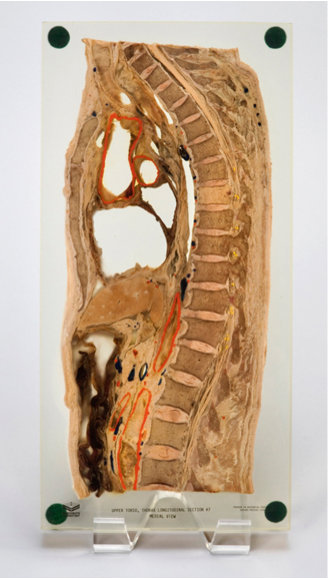
Sagittal Section of Human Spine Preserved in Arcrylic
In the late 19th century, German anatomists pioneered a new method that revealed bodily structures and their spatial relationships in amazing detail. They froze cadavers and cut them into very thin cross-sections.
This unnatural and abstract way to see a body became newly relevant for physicians with the advent of computerized tomography (CT) scans and magnetic resonance imaging (MRI). By the 1970s, anatomical preparators had begun preserving thick cross-sections of donated cadavers in acrylic slabs. Students could hold them side by side with radiological images for comparison.
Prepared this way, the cadaver could survive decades, if not hundreds of years, serving as an enduring teaching tool for human anatomy. Even though this is an abstract way to encounter a body, the slices are thick enough to convey a strong sense of corporeality.
Sarcosote Laboratory and Program in Anatomical Education, Harvard Medical School, 1970s
Program in Medical Education, Harvard Medical School